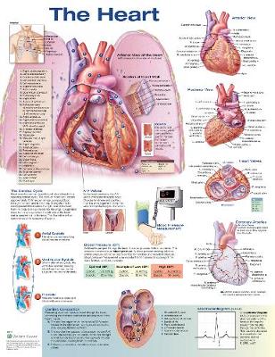
One of Anatomical Chart Company&;s most popular charts,
The Heart, Second Edition features a large central image that shows the heart sitting on the diaphragm with the suggestion of the lungs and ribs behind it. Cutaways on the anterior wall of the heart show the interior structures, and additional images present normal anatomy, the cardiac cycle, blood pressure measurement, cardiac conduction, and more. All features are clearly labeled.
Normal heart anatomy featured:
- Posterior and anterior views of the heart
- Detail of a section of heart wall
- Heart valves
- Coronary arteries
- Thorax, showing the location of the heart
Also included are the following diagrams and definitions:
- The cardiac cycle
- A-V valves
- Blood pressure (BP) and measurement
- Electrocardiogram (ECG)
- Cardiac conduction
Medical illustrations by Marcelo Oliver, MFA and Lik Kwong, MFA, in consultation with Leonard S. Lilly, MD, Harvard Medical School, Brigham and Women's Hospital, Boston, Massachusetts
20" x 26" heavy paper laminated with grommets at top corners ISBN 9781496369628
One of Anatomical Chart Company&;s most popular charts,
The Heart, Second Edition features a large central image that shows the heart sitting on the diaphragm with the suggestion of the lungs and ribs behind it. Cutaways on the anterior wall of the heart show the interior structures, and additional images present normal anatomy, the cardiac cycle, blood pressure measurement, cardiac conduction, and more. All features are clearly labeled.
Medical illustrations by Marcelo Oliver, MFA and Lik Kwong, MFA, in consultation with Leonard S. Lilly, MD, Harvard Medical School, Brigham and Women's Hospital, Boston, Massachusetts
20" x 26" heavy paper laminated with grommets at top corners ISBN 9781496369628